Restorative dentistry in animals functions much like dental fillings in humans. Although pets can develop cavities, they are far less common than in people. Instead, veterinarians more frequently address other enamel abnormalities using dentin bonding and composite restoration techniques. Tooth fractures are especially prevalent in both dogs and cats and may result from trauma—such as being struck by a hard object—or from chewing items that are excessively rigid. Any tooth can break, but canine teeth and, in dogs, the upper fourth premolars are among the most frequently affected.
Fractured teeth are generally grouped into two main categories: those that expose the pulp within the root canal (complicated crown fractures) and those that do not reach the pulp but still reveal dentin (uncomplicated crown fractures). Both types require treatment, though the appropriate therapy depends on the severity of the injury. When the root canal is exposed and left untreated, bacteria can infiltrate the pulp, causing the tooth to lose vitality and become a reservoir for infection. Over time, the infection may extend to the bone around the tooth roots and, eventually, bacteria may spread through the bloodstream to internal organs.
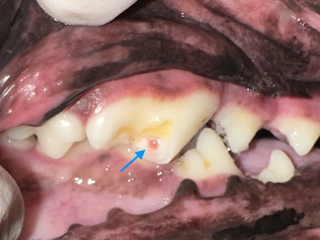
A complicated crown fracture that exposes the pulp. The pulp appears pink (blue arrow) indicating the fracture is recent.
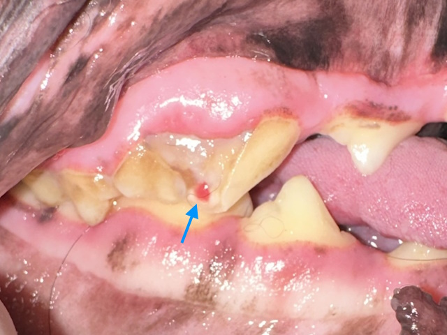
A complicated crown-root fracture that exposes the pulp. The pulp appears pink (blue arrow) indicating the fracture is recent. The fracture extends below the gumline.
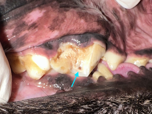
A complicated crown fracture that exposes the pulp. The pulp appears brown (blue arrow), indicating an old fracture that caused pulp infection and necrosis.
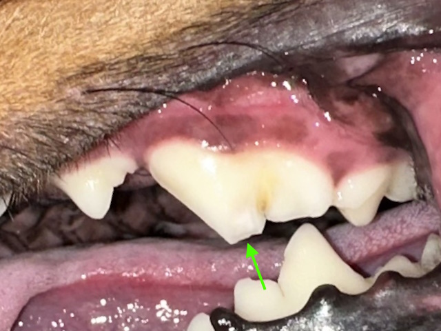
An uncomplicated crown fracture that caused exposure of dentin (green arrow), but not the pulp.
For teeth with direct pulp exposure, only two treatment paths exist—root canal therapy or extraction—and simply ignoring the problem is not an option. Root canal therapy is generally considered the preferred approach when the damaged tooth remains structurally healthy, as it preserves the tooth’s function. Extraction, on the other hand, is typically reserved for situations in which root canal therapy is not feasible. Removing strategic teeth such as canine teeth, the upper fourth premolars, or the lower first molars is avoided when possible because these teeth have large, complex roots that make extraction more invasive and because their loss can impair chewing function and even lead to orthodontic complications like lip entrapment or gum trauma.
Uncomplicated crown fractures require a different strategy. These injuries, which are especially common in large-breed dogs, often involve the major chewing teeth—the maxillary fourth premolars and mandibular first molars. Even without pulp exposure, dentin is sensitive, and uncovered dentin causes significant discomfort similar to dental sensitivity in people. Some of these teeth will eventually die and become infected; in fact, recent research has found that nearly one-quarter of such fractures lead to non-vital teeth. For this reason, dental radiographs are recommended to assess the health of the affected tooth. If the tooth is dead, treatment involves either root canal therapy or extraction. If the tooth is still alive, applying a bonded sealant can reduce pain, seal off bacterial pathways, and smooth the surface to help prevent periodontal disease—an approach that has shown excellent success.
Other conditions, such as enamel hypoplasia and true dental caries, can also necessitate restorative procedures. Enamel hypoplasia occurs when the enamel fails to form correctly, leaving the tooth rough, pitted, and yellowed. Pets with this condition may experience sensitivity, increased tartar buildup, periodontal disease, infection, or abnormal root development. Treatment options depend on the severity of the defect and the functional importance of the tooth and may include dental imaging, thorough cleaning, sealants, composite restorations, crowns, or extraction.
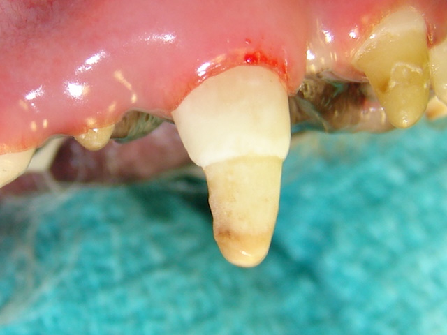
Enamel hypoplasia affecting nearly half of the canine tooth crown away from the gumline.
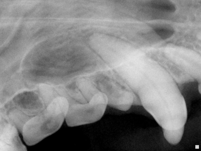
Dental radiograph of this canine tooth does not reveal any root or root tip abnormalities.
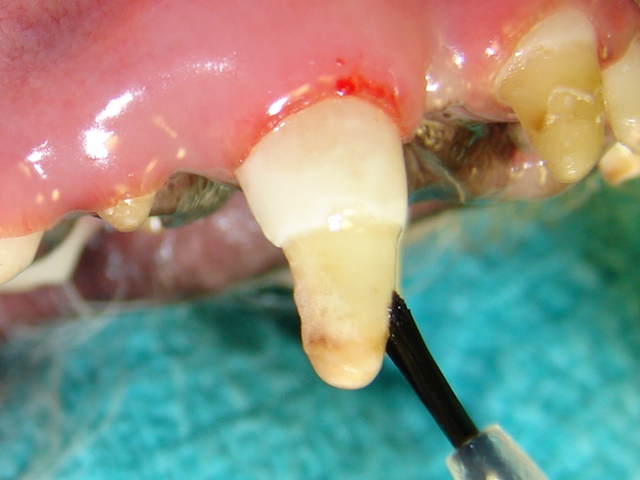
A dentin bonding agent is being applied to the exposed dentin to seal off the dentinal tubules.
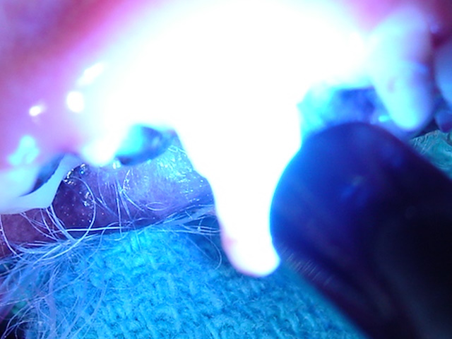
Dentin bonding agent on the tooth is exposed to ultraviolet light to trigger a reaction that causes the liquid resin material to polymerize and solidify.
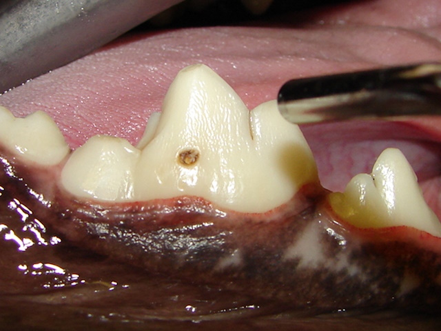
A small enamel defect is present on the outside surface of the lower first molar tooth in a dog.
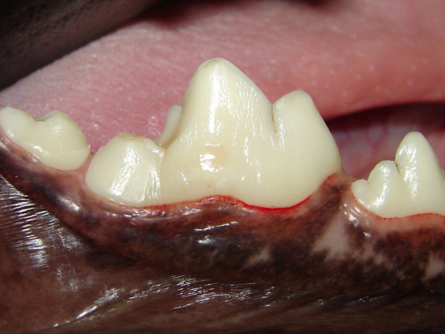
The defect is prepared and filled with composite filling material.
Bacterial cavities—known as caries—are uncommon in pets but can still appear, most often on the flat surfaces of molars. They typically present as dark brown or black areas but must be distinguished from staining caused by wear. Treatment mirrors that of human dentistry, although in cases where the tooth structure is significantly compromised, extraction may be the most appropriate choice.
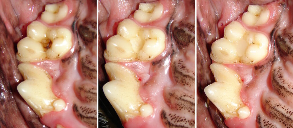
A cavity on the chewing surface of the upper first molar in a dog is cleaned and the defect is filled with a composite filling material.
A cavity on the chewing surface of the upper first molar in a dog is seen (blue arrows).
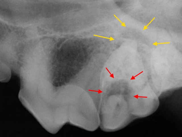
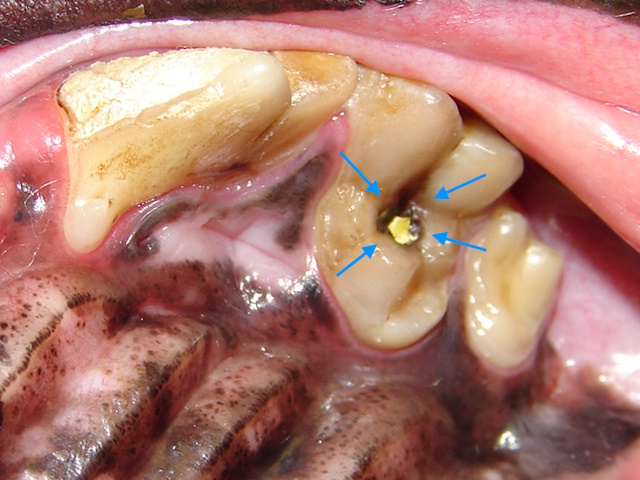
This cavity has created a deep defect and infection in the crown (red arrows) which already spread to the pulp and the bone around the root tip (yellow arrows).
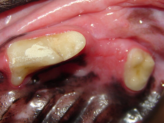
This tooth was deemed structurally irreparable, and it was extracted to get rid of the infection. Extraction site healed well in a month.
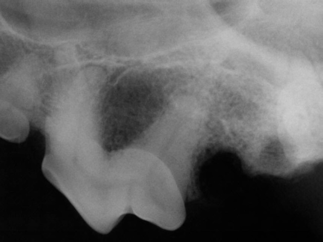
Post-extraction dental radiograph shows complete extraction of all the roots.