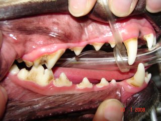
In stage 1 (gingivitis), the gum lining the teeth is inflamed and swollen. There is plaque covering the tooth surface, mostly closer to the gum.
In stage 2 (early periodontitis), the inflammation of the gums are worse. The teeth are covered with significant amount of tartar. The pet’s mouth is painful.
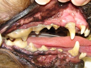
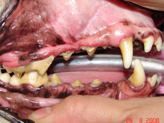
In stage III (established periodontitis) the gum lining starts receding. The bone loss is getting worse and roots are being exposed.
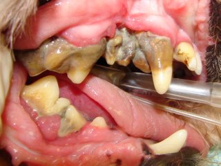
In stage IV (severe periodontitis) there is severe gum recession, bone loss and root exposure. The periodontal supporting structures are almost entirely destroyed and the teeth are mobile. Bacteria may be spreading throughout the entire body via the bloodstream and may damage the kidneys, liver and heart.
While periodontal disease is irreversible, treatment at any point in the disease process will stop or slow this progression. Without treatment, the end stage of this disease is tooth loss; however the disease has likely caused problems well before this. Bacteria from infected gums might gain access to the systemic circulation. Consequences of bacteria circulating in the main blood stream include kidney infections, liver abscesses, infectious arthritis, and birth problem in puppies. These bacteria may also become attached to heart valves and cause endocarditis which results in an intermittent infection and strokes. Clinical signs of gingivitis are swelling, a gingival color change from pink to red, bleeding gums, significant tartar, and bad breath. Clinical signs of periodontitis include the above plus gum recession, difficulty eating, and increased tooth mobility.
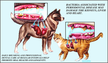
Periodontal disease can cause infections in the kidneys, liver, heart, joints and other distant sites in your pet’s body.
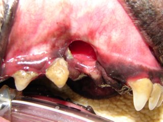
There are two common consequences to severe periodontal disease. The first and most common consequence is an oral-nasal fistula. The roots of the upper canine teeth (fangs) are next to the nasal cavity and separated from it by only a thin sheet of bone. Periodontal disease destroys this thin bone resulting in communication between the oral and nasal cavities. The bacteria, food particles, and other oral debris will enter this area and cause an infection in the nasal cavity. Signs are chronic nasal discharge, sneezing, and occasionally loss of appetite and bad breath. This is diagnosed by the veterinarian introducing a probe into the area under general anesthesia. The treatment in most cases requires surgical intervention.

Root of the upper canine tooth is separated from the nasal cavity with a very thin layer of bone which can be destroyed by periodontal disease resulting in communication between the oral and nasal cavities.
Inappropriate or no closure of the extraction site after upper canine tooth extraction resulted in a big oronasal fistula in this dog.
In addition, periodontal disease will weaken the bone in the affected areas. In the lower jawbone it can weaken to the point of causing a pathologic fracture. The fractures occur most commonly due to mild trauma (like jumping off a couch); however, some dogs have broken their jaw while eating. The fixation and healing of these fractures is usually very difficult due to the lack of remaining healthy bone.
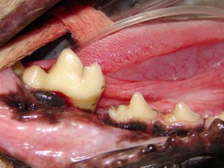
This dog had a very mobile lower jaw bone on the right side and he was painful on manipulation of the lower jaw.
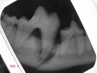
Dental x-ray reveals the significant bone loss around the mesial root and fractured jaw bone. Severe bone loss around the mesial root of this lower first molar tooth is caused by periodontal disease which eventually led to jaw fracture between the fourth premolar tooth and first molar tooth.
Keep in mind, this very common and severe disease process can be prevented or slowed with routine professional cleanings and homecare. Please note that the bacteria that cause this disease are located under the gum line. Anesthesia free “cleanings” DO NOT address this area and are of little to no therapeutic value. Please consult with us or with your veterinarian as to the proper therapy for your pet.