Endodontics is the branch of dentistry that deals with the etiology, diagnosis, treatment, and prevention of diseases and injuries of the pulp,nerve and blood vessels in the center of a tooth. Endodontic therapy is colloquially known as “root canal therapy.” Put simply, a root canal therapy involves removing the infected pulp tissue of a dead tooth, then placing inert materials inside the root canal to make the affected tooth an infection and pain free tool for eating and playing again.
A fractured upper incisor’s pulp chamber and root canal are being cleaned with an endodontic file.
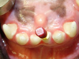
The incisor’s pulp chamber and root canal are filled with an inert material and a sealer cement to prevent future bacterial leakage into the canal space.
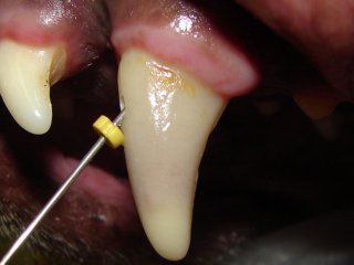
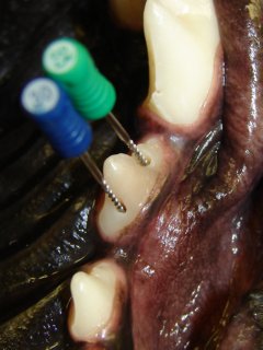
A canine tooth with purplish color change is being cleaned out with an endodontic file.
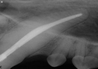
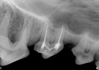
Canine tooth’s root canal space is filled with sealer cement and an inert material.
Upper third premolar tooth’s inside is being cleaned with endodontic files.
Third premolar tooth is filled with an inert material.
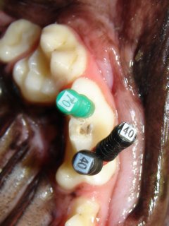
Upper fourth premolar tooth has three roots. Three endodontic files are used to clean out all these roots.
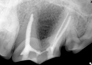
The roots are filled with an inert material.
Broken or fractured teeth are very common in dogs and cats. The most common causes of broken teeth are abnormal chewing and biting behaviors (chewing on rocks, cow hooves, animal bones, hard nylon toys, ice cubes, etc), trauma (hit-by-car, falling, etc), and rough play. The most commonly affected teeth are incisors, canine teeth and upper fourth premolar teeth. Common signs of tooth fractures and resultant endodontic disease include drooling more than usual, pawing at face, rubbing face on carpet or eating on the other side of the mouth. Other less common signs are teary eyes, greater tartar buildup on one side of the mouth, a gray or purple discoloration of a tooth, and sneezing.
A common phrase we hear from pet owners is: “It does not seem to be bothering him.” This “appears” to be true because pet owners may notice no change in their pet’s behavior to alert them. However, pets feel pain too. A dog or a cat will not stop eating because he has a sore tooth. The biggest sign of endodontic disease is no sign at all. Therefore, it can be very hard for an owner to notice the problem unless they routinely brush their pet’s teeth and see the broken or discolored tooth.
Fractures of the teeth open portals for oral bacteria to invade pulp tissue. These portals can be opened by direct exposure of the pulp chamber or exposure of the dentinal tubules (microscopic canals in the dentin that run from the enamel-dentin junction to the pulp chamber). As the bacteria reaches the pulp tissue, infection starts. Presence of bacteria in the root canal system causes an inflammatory reaction in the tissues around the root tip. This inflammatory reaction serves two purposes – one is to try to remove the bacteria, and the other is to prevent microbial invasion into the tissues around the root tip. Removal of bacteria from the root canal system is usually unattainable due to lack of blood supply in the dying pulp tissue. The bacteria will spread to the root tip and try to get out of the root canal into the tissues (jaw bone) around the root tip. However, the immune system cells will constantly fight the infection in this area and will keep it under control. The root canal system and the root tip area will be chronically infected. This chronic infection represents a ‘balance’ between the bacteria in the tooth and the body’s response. Once this ‘balance’ is disturbed, an acute inflammatory reaction will develop with severe symptoms. This may occur spontaneously as a result of different factors produced locally at the site of the ongoing inflammatory reaction, or as a result of an immune reaction. The symptoms seen will vary depending on the course that the inflammation follows. It can be seen as further intensification of the inflammation (acute sharp painful episode), abscess formation (primary apical abscess), development of a sinus tract (chronic apical abscess), spreading of the infection through bone and/or soft tissues (cellulitis), and cyst formation. Healing will only occur if no further irritation occurs to sustain the reaction and if there are no bacteria within the canal – such as after root canal therapy has been completed.
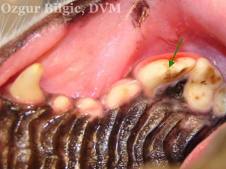
Uncomplicated crown fracture of upper fourth premolar tooth. The pulp chamber is not directly exposed but the dentin is; hence, dentinal tubules are exposed to oral cavity.
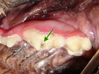
Complicated crown fracture of upper fourth premolar tooth. The pulp chamber is directly exposed to oral cavity. The pulp appears black as a result of pulp tissue death due to chronic infection.
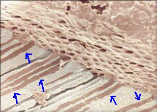
In this microscopic image dentinal tubules are seen (blue arrows) running across the width of the dentin.
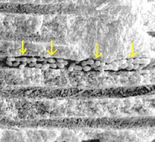
Electron microscopic view reveals bacteria packed dentinal tubule (yellow arrows).
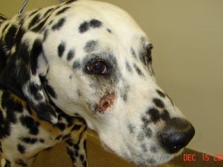
Chronic apical abscess resulted in formation of a draining sinus tract below the eye.
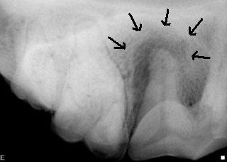
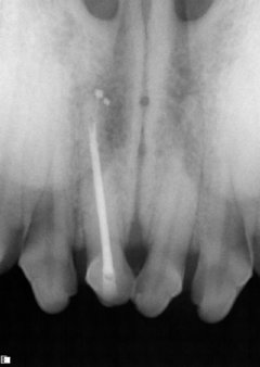
Dental radiographs of the dog on the left revealed a tooth fracture and root abscess associated with upper fourth premolar tooth.
Antibiotics will help control the infection temporarily, but the problem recurs after discontinuing the medication. The infection will persist until the source of the infection, the infected pulp, is removed either by extracting the tooth or by root canal therapy. The age of the patient, extent of infection, amount of tooth or root damage, periodontal health of the tooth, duration of pulp exposure and importance of the tooth will help decide which treatment is best for the patient.