Head and neck surgery in dogs and cats includes a range of procedures used to treat conditions affecting the face, salivary glands, ears, eyes, nasal passages and soft tissues of the neck. These surgeries often require advanced surgical skills due to the dense concentration of nerves, blood vessels, and other vital structures in this region. Also, some procedures will require excision of large amount of tissue followed by reconstructive surgery. The primary goals of head and neck surgery are to relieve pain, remove disease, and preserve normal function while maintaining the animal’s quality of life.
Soft Tissue Surgery of the Face and Neck
Soft tissue surgery of the face and neck addresses a variety of conditions, including masses, cysts, abscesses, and enlarged lymph nodes. Neoplastic masses on the face, head, and neck may require wide margin excision and skin reconstruction to completely rid of the cancerous tissue. Lymph node biopsies or excisions are commonly performed to diagnose infections, inflammatory diseases, or cancer. Surgical exploration of the neck may also be required after bite wounds or penetrating injuries to assess damage to deeper structures and prevent complications such as infection or bleeding. Severe lacerating trauma may also require reconstruction of the facial soft tissues.
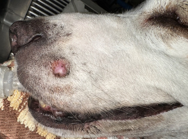
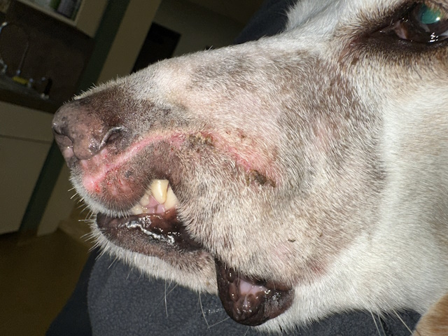
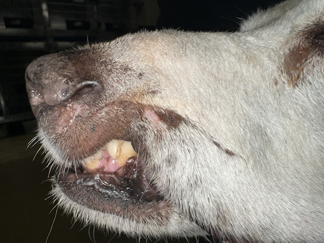
A dog with a constantly growing apocrine ductular carcinoma on his left upper lip. The mass was penetrating the full thickness of the upper lip,all the way to the oral mucosa inside the mouth.
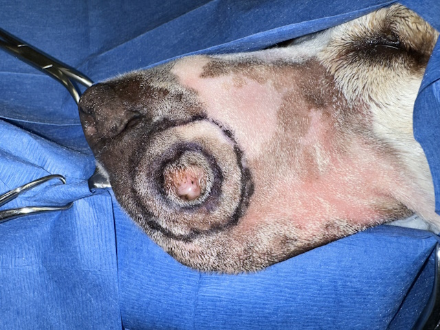
The surgical field was draped and prepared for surgery. The extent of the mass and a 2 cm margin excision line was drawn on the skin with a marker.
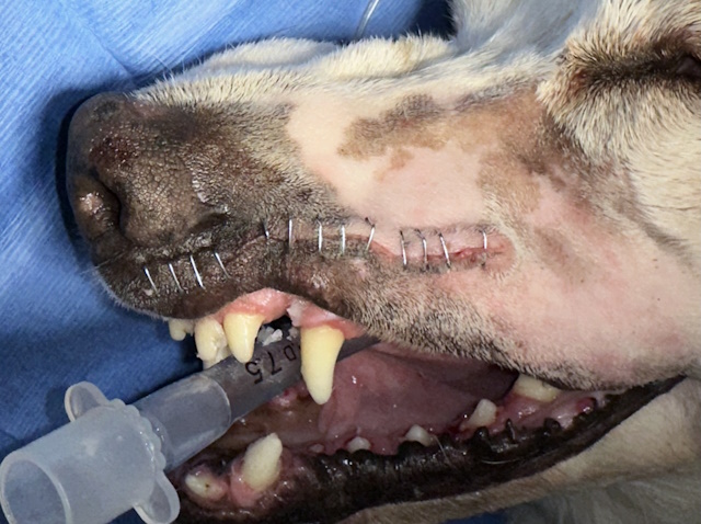
Full thickness excision of the mass was performed. Subcutaneous tissues were closed with absorbable suture material. Skin was closed with stainless steel staples.
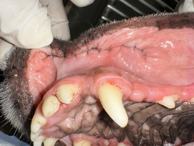
Oral mucosa was closed using an absorbable suture material using a buried knots technique.
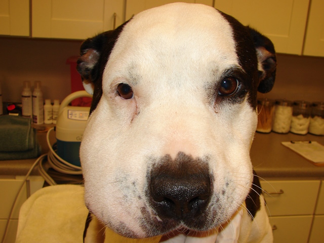
Recheck of the surgical site 10 days after surgery during staple removal. The area has healed well.
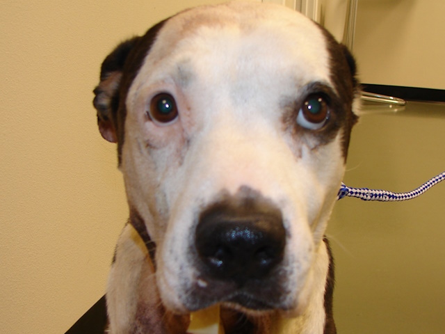
Recheck of the surgical site 6 months after surgery. No sign of tumor recurrence is seen.
A different dog with a large abscess on the right side of his face. This required abscess drainage and surgical debridement of the abscess pocket followed by multiple drains.
This picture is taken about a week after treatment. Notice the significant improvement in patient’s face.
Salivary Gland Surgery
One of the most commonly performedhead and neck procedures in dogs and cats is the treatment of salivary gland disorders. Salivary mucoceles occur when saliva leaks from a damaged salivary gland or duct and accumulates beneath the skin, forming a soft swelling. Surgical treatment typically involves removal of the affected salivary gland and its associated duct to prevent recurrence. In some cases, salivary gland surgery is also performed to remove tumors or address chronic infections that do not respond to medical treatment.
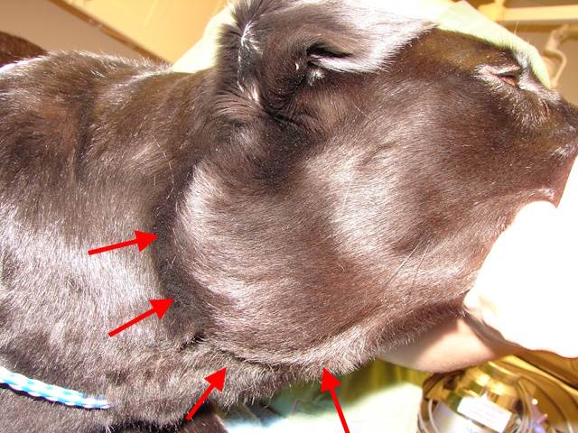
A cervical salivary mucocele on the right side of the neck of this dog.
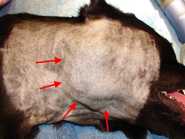
Mucocele can be better appreciated after this area was clipped as preparation for surgery.
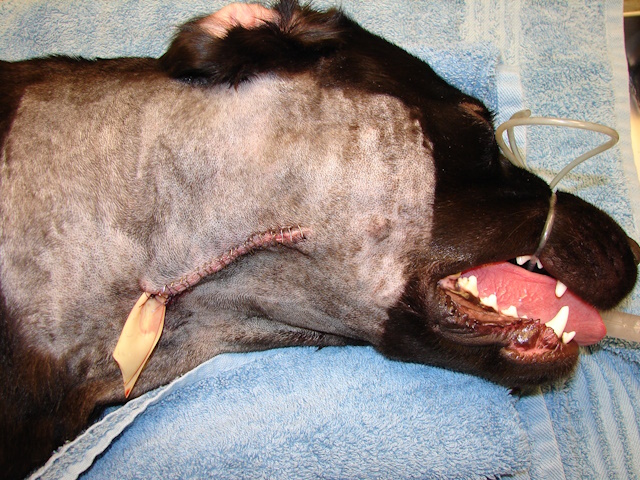
Surgical removal of the affected salivary gland complex (mandibular and sublingual) was performed, and a drain tube was placed.
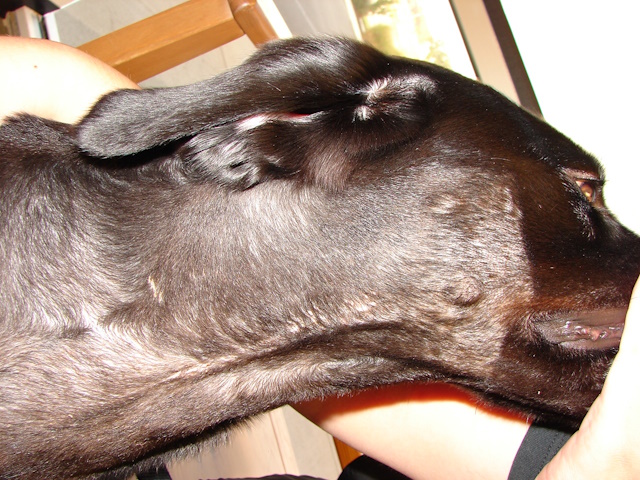
This picture is taken 8 days after surgery. Salivary mucocele is gone, and the surgical site has healed well.
Special Considerations in Head and Neck Surgery
Surgeries in the head and neck region require careful planning due to the close proximity of critical structures. Protecting major blood vessels and nerves is essential, as damage can result in serious complications. Postoperative swelling and discomfort must be closely monitored, and effective pain management is an important part of recovery. Because these surgeries can affect eating, grooming, or movement of the head, attentive postoperative care helps ensure a smooth healing process.